PAI is provided with a demonstration neural network architecture designed for multichannel segmentation. This architecture expects one or more 3D input series and generates segmentation results with one or more segments/VOIs.
The initial application of this architecture was for a case called Tumor Detection. A trained model Tumor Detection is available in the Segmentation tool when PAI is licensed, using the Multichannel Segmentation architecture. It is based on the MICCAI Brain Tumor Segmentation (BraTS) Challenge: http://braintumorsegmentation.org. BraTS utilizes multi-institutional pre-operative MRI scans and focuses on the segmentation of intrinsically heterogeneous (in appearance, shape, and histology) brain glioma tumors.
Training and testing of the Tumor Detection model in PAI was performed using the data from the 2020 BraTS Challenge containing 369 samples. Each sample consists of four MR images (native T1, post-Gd-contrast T1-weighted, T2 FLAIR, T2-weighted) and one image containing three reference segments as label numbers.
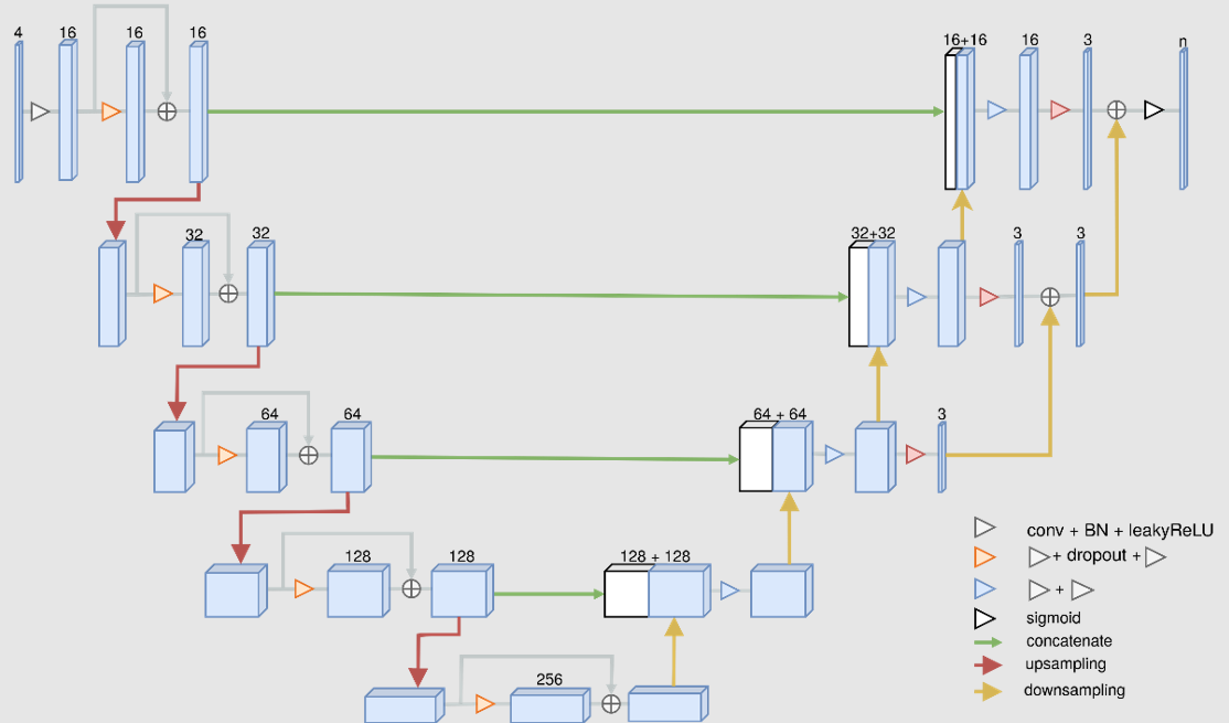
The Multichannel Segmentation architecture is a modified version of the convolutional neural network U-Net:
The output of the Tumor Detection model is a label image with three segments (label 1: non-enhancing tumor; label 2: peritumoral edema; label 4: Gd-enhancing tumor).
A second experimental model based on the Multichannel Segmentation architecture is included with PMOD 4.203. This model is called Rat Brain Dopaminergic PET. It requires early and late average images derived from PET with tracers such as [11C]-raclopride and [11C]-methylphenidate, and the output is a label image with three segments (label 1: cerebellum; label 2: left striatum; label 3: right striatum). The model was trained using 382 rat brain dopaminergic system dynamic PET datasets. A description of the preparation of the data and process to train and evaluate the model is included later in this documentation as a case study.
BraTS Reference:
Bakas, Spyridon & Reyes Jan. (2019). Identifying the Best Machine Learning Algorithms for Brain Tumor Segmentation, Progression Assessment, and Overall Survival Prediction in the BRATS Challenge. https://arxiv.org/pdf/1811.02629.pdf