The signal of a dynamic PET measurement represents the averaged activity in the image pixels at acquisition times t starting at tracer injection. It is described by the operational equation

This equation means that the activity concentration CPET measured by PET in a certain tissue volume is composed of two contributions:
▪tracer which has been extracted into tissue, where it has an instantaneous concentration of CTissue(t).
▪tracer which is circulating within the blood, with a concentration CBlood(t).
Hereby it is assumed, that the fractional volume vB is composed of small capillaries (2%-5%) filled with whole blood, and that the fraction (1-vB) represents tissue. The blood activity CBlood must be measured during the acquisition. In tissue, the tracer may be present in different spaces or forms (eg. free, specifically bound, non-specifically bound), which are described by the compartments of the model. All compartments contribute to the tissue signal, so that it is modeled by the expression
whereby
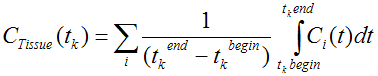
▪CTissue(tk) represents the average concentration of tracer in tissue during the acquisition k which starts at tkbegin and and lasts until tkend
▪Ci(t) represents tracer concentration in compartment i at time t . These expected concentrations are calculated from the differential equations using the current model parameters and the plasma input curve(s).
The operational equations used for other than compartment models are specified in the PKIN Model Reference section.