When a compartment system has equilibrated, the total distribution volume can be calculated easily as the ratio of tracer concentration in tissue to the metabolite-corrected plasma concentration with a single static scan. It has been found that the time required to reach equilibrium can be shortened by an optimized tracer delivery. A setup which does not require a sophisticated tracer delivery system is to apply an initial bolus and continue with a constant infusion.
Carson et al. [1] have developed a method to optimize the ratio between the activity given as the initial bolus and the activity level of the subsequent infusion for quickly reaching an equilibrium (Appendix B in [1]). This method is implemented as a tissue model in PKIN. The current blood model Bolus/Infusion is only a visualization model and should not be used for fitting measured blood data. It allows entering the Kbol value found with the tissue model optimization and inspecting the plasma activity curve under the bolus/infusion regime.
Operational Model Curve
For a bolus and infusion (B/I) experiment the expected input function can be derived from the input curve CP(t)of a bolus-only experiment by
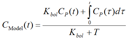
Kbol [min] defines the dose of the bolus expressed as minutes of infusion, and T is the total duration of the infusion (end of last frame).
Parameter Fitting
This model cannot be used for data fitting. It assumes the plasma activity curve of a bolus experiment has been loaded and visualizes the expected input curve if Kbol [min] equivalents of the infusion are applied as an initial bolus. The criterion of fast equilibration is that the model curve gets soon constant and remains so.
Reference
1.Carson RE, Channing MA, Blasberg RG, Dunn BB, Cohen RM, Rice KC, Herscovitch P: Comparison of bolus and infusion methods for receptor quantitation: application to [18F]cyclofoxy and positron emission tomography. J Cereb Blood Flow Metab 1993, 13(1):24-42. DOI