To start parcellation of a brain MR series, select the Brain Parcellation tab of PNEURO and activate the MR button in the lower right of the FUNCTIONAL page to move to the ANATOMICAL T1 MR page. The image should be T1-weighted, cover the entire brain and have a pixel size of about 1mm. After loading, the image should appear in radiological HFS orientation.
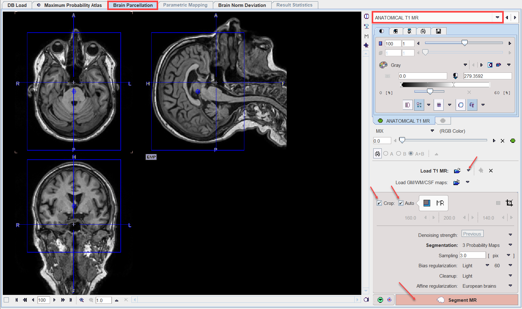
MR Image Cropping
The MR image is the basis for the parcellation. Experience has shown that problems may occur if the MR field-of-view is much larger than the brain as occurs for instance with sagittal MR acquisitions. Therefore, please use the Crop facility to limit the MR data set to the relevant portion with skull and brain.
MR Image Segmentation
The MR image needs to be segmented into gray matter (GM), white matter (WM) and cerebrospinal fluid (CSF). The settings that influence the algorithm are described above. The actual segmentation is started with the Segment MR action button. Note that the denoising and segmentation process may take several minutes.