The result of matching is shown on the MATCHED PET page. Please verify that matching was successful by evaluating the alignment in different parts of the brain. Particularly helpful to do so is to interactively drag the fusion balance left/right, and to enable contour outlines.
If the match is not satisfactory, there are two options to rectify the situation:
1.Return to the previous page, change the sampling and smoothing parameters and try the automatic matching again, or
2.Activate the Adjust matching button and shift/rotate the PET image interactively by dragging the handles in the image or entering offsets/angles on the Move/Rotate tabs. Finally the transformation needs to by applied with the ![]() button.
button.
Normalization
The next step is to spatially normalize the subject images. There are three options as illustrated below.
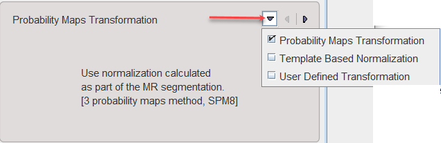
Probability Maps Transformation |
Uses the normalization resulting from the GM/WM/CSF MR segmentation procedure. |
Template Based Normalization |
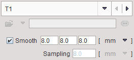
Performs an SPM5-type normalization between the subject MR and the T1 atlas template image with the usual options |
User Defined Transformation |
For loading and applying a normalization transformation which has previously been calculated for the MR image and saved. |
Please activate the Normalize action button to proceed.