The layout of the pages for the Brain VOIs calculation are illustrated below using the N30R83 Maximum Probability Atlas.
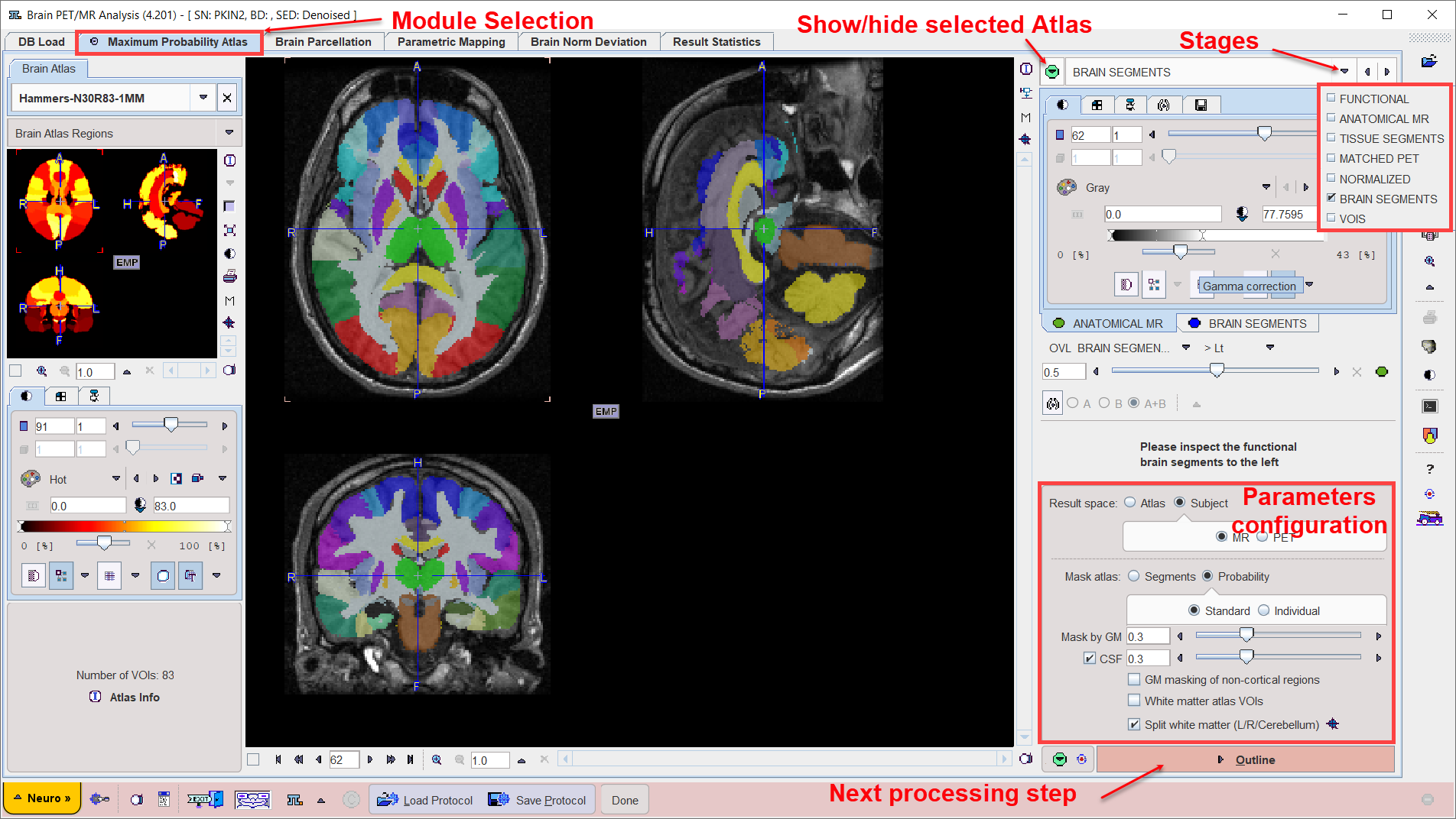
The main part is used for displaying the images with some overlay information. The control area is located to the right, and an optional part related to templates is shown to the left. The template area is shown/hidden by the indicated green button in the upper right.
Step-Wise Processing
Data processing is consistently organized by a stepwise progression towards the end result. On each processing stage the user has to take some action such as data loading, alignment inspection or parameter configuration, and then start the next processing step with the red action button located in the lower right. As soon as the result is calculated, it will be shown on a new page representing the new processing stage. The cascade of stages is available by the selection area in the upper right.
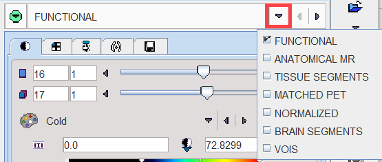
It conveniently allows inspecting the results of prior stages without initiating any calculations. To repeat a calculation with modified parameters, the action button in the lower right has to be activated again on the actual and all following pages.
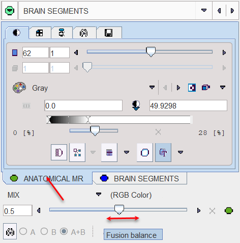
Fusion Image Display
The display of the images is controlled in the upper right. In many cases more than one image contributes to the display. In these cases the tab corresponding to an image has to be first activated, before its color table or the color thresholds can be modified. The fusion control section is located below the image control tabs. In the configuration illustrated below the colors of both images are mixed, whereby the weighting can be changed with the slider.
Convenience Buttons
Next to the action button in the lower right is an area with two buttons
![]()
offering the following functions:
|
Hide the parameters panel to free some space in the user interface. With the panel hidden, the icon changes to |
|
Reset the parameters on the page to their default values. If the button in the taskbar to the right is activated, the defaults are reset on all pages. |
Configurations
The PNEURO tool can be configured according to user preferences in a dialog window as illustrated below.
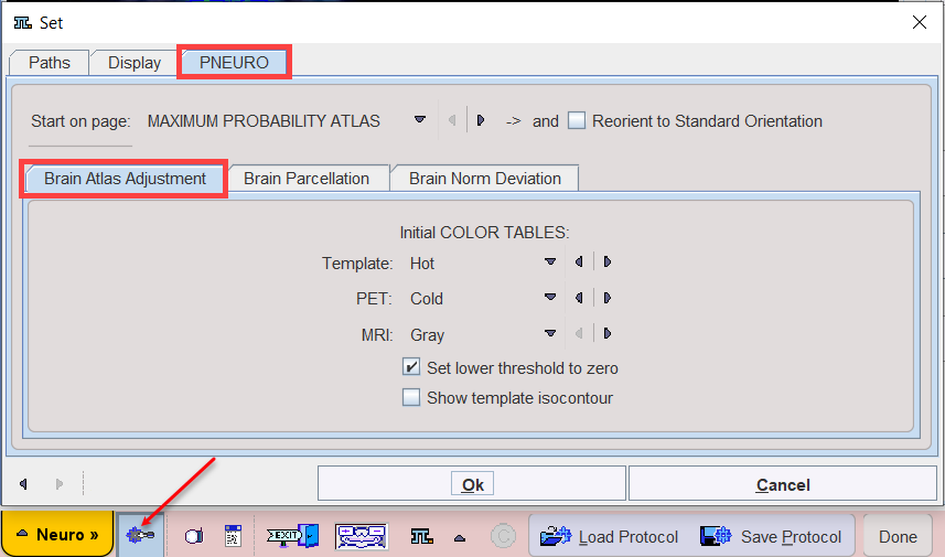
The common configurations are available on the Paths and Display tabs, and in the upper part of the PNEURO tab. Note the Reorient to Standard Orientation box. If it is checked, PNEURO tries to orient the brain images such that they appear in the radiological Head First Supine (HFS) order with subject left on the image right. For instance, MR images acquired in sagittal orientation will automatically be reformatted and presented with axial slices. The correct HFS orientation of the data after loading is important for the automatic procedures to work properly.
Further Information
This guide is focused on the brain analysis functionality. Please refer to the PMOD Base Functionality Guide for details about general functions such as data loading, image display, and VOI definition.