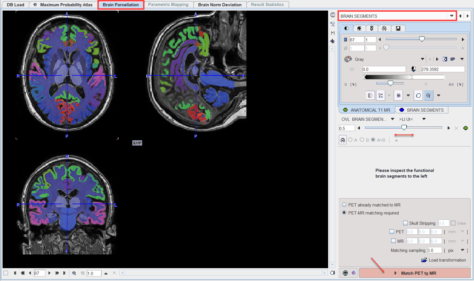
The BRAIN SEGMENTS page shows the parcellation result as an overlay onto the MR image.

The BRAIN SEGMENTS image represents a label atlas of the brain structures identified by the segmentation algorithm. Please check the alignment of the segments with the subject image using the fusion slider. In the case of a mismatch, try changing the GM segment definition on the (previous) TISSUE SEGMENTS page and/or the number of subjects included and repeat parcellation. Alternatively, the structure definitions can be adjusted manually after the outlining step.
PET to MR Matching
The next step consists of rigidly matching the averaged PET image to the MR image. The matching options are in the lower right and described above.
Please activate the Match PET to MR action button to start matching.