PET images are inherently affected by the partial-volume effect. This means that the measured tracer activity concentration is not accurate due to the relatively low image resolution and the limited tissue sampling. The low spatial resolution of the PET system causes a blurring of the image, so that high activities (from a hot lesion) are spread to the surrounding as illustrated below. This effect is called spill-out. The same effect also causes a spill-in of background activity into the volume of interest.
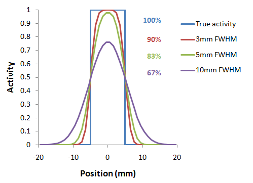
As a consequence, hot lesions tend to appear less aggressive (reduced maximum) but bigger (spreading) than they are in reality.
Spill-in and spill-out depend on the geometry of the objects, the activity distribution of the tracer, and on the resolution of the scanner which may vary across the imaging field-of-view. Therefore, practical correction approaches have to assume certain conditions and can only be approximate. For a nice overview of the topic please refer to the publication of Soret et al. [1].
Partial-volume correction in PNEURO is accessed on the VOIs page of either the Maximum Probability Atlas or Brain Parcellation workflows:
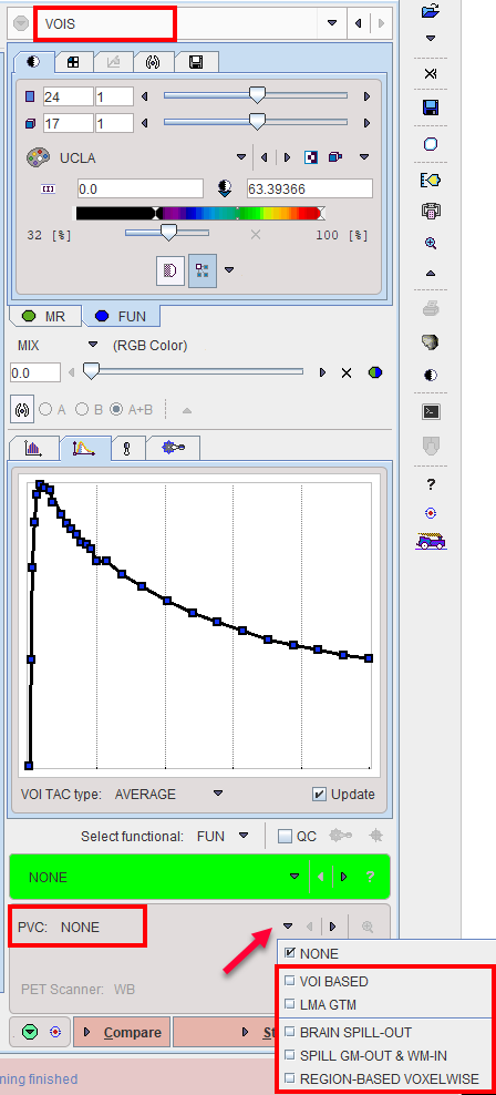
The selection and configuration of the methods available is described in more detail in the Workflow sections of this document.