After successful rigid matching and normalization the mapping between the different image spaces is established:
▪the normalization transform maps the Anatomical MR to the atlas space;
▪the rigid transform maps the Input to the Anatomical space;
▪the rigid transform combined with the normalization transform maps the Input to the atlas space.
As all the transformations can be inverted, the atlas space can also be mapped to the Input and the Anatomical MR image space. Consequently, the brain structures which are defined in the atlas space can be mapped to the Input and Anatomical MR subject space and shown in the overlay.
Calculate the Brain Segments
Activate the Segment Brain action button at the right bottom of the NORMALIZED layout to prepare the images in all possible spaces. As a result, the original Anatomical image is overlaid with an image of the transformed atlas regions called Brain Segments. Please perform a final validation of the region placement. As mentioned before, a certain degree of deviation can't be avoided and needs to be handled by adjusting the generated brain VOIs.
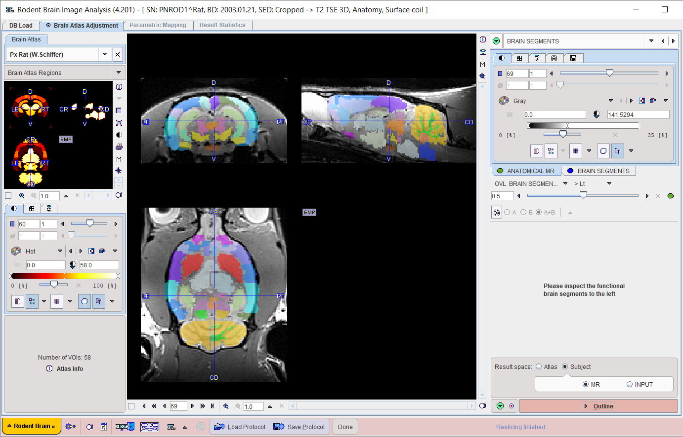
Result Space Definition
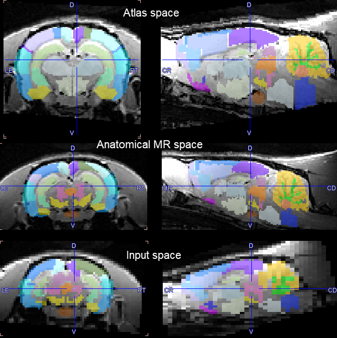
There are three image spaces where the results can be generated and the statistics calculated:
1.Atlas space: The Input and the Anatomical images are transformed to the space of the atlas. This option is preferable for pooling the resulting images (functional image, parametric map) of a group of subjects and performing an analysis such as SPM.
2.Subject/MR: The Input image and the brain VOIs are transformed to the Anatomical space. This option can be preferable for visualization purposes if Anatomical has better resolution than the Input.
3.Subject/INPUT: The Anatomical image and the brain VOIs are transformed to the Input space. This option has the advantage that the statistics are calculated on the original Input data, whereas in the other choices the data has been resampled.
The information visualized on the page is updated as soon as the configuration is changed. The illustration below shows the effec when selecting the different spaces. Clearly, the PET Input series has the lowest resolution so that the resliced Anatomical image appears heavily pixelated.
Brain Structure Outlining
Once the result space has been specified, the brain structures are fully defined and can be outlined to create contour VOIs. This process is started with the Outline action button.