This is the model which supports the quantitative data analysis for the classical autoradiography with 14C-deoxyglucose (DG) [1]. In fact it is this model from which the PET FDG autoradiography model is derived from, and both have the same underlying assumptions and equations.
In summary, an autoradiographic experiment is performed as follows:
1.The 14C-labeled deoxyglucose is injected.
2.Blood is sampled and counted until the end of the experiment.
3.The glucose concentration in plasma is measured for one sample.
4.After 50 minutes the deoxyglucose has been trapped and the animal is sacrificed.
5.The brain is isolated, then frozen, and sectioned into very thin slices.
6.The slices are put onto a flat support and mounted into a radioactivity counter together with reference sheets of known activity concentration.
7.The radioactivity is counted during several days.
The result is a set of images either on a conventional film or as a digital file in one of the popular graphic formats. These images can be turned into radioactivity units by a translation table which needs to be obtained from the reference sheets.
Acquisition and Data Requirements
Image Data |
A data set representing the autoradiographical slices in arbitrary units. Select 1/1 as the loading units as the values are transformed into nCi/g during the model calculations. An appropriate translation table must be derived from the image representation of the reference sheets and supplied as the TAC1 curve in the model preprocessing panel. Note that the duration of the acquisition must be specified as the time from injection until sacrificing the animal. |
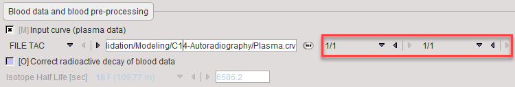
Blood Data |
Plasma activity of blood sampled at a peripheral artery from the time of injection until sacrificing the animal. Important: Select 1/1 as the units and ensure that the values in the data file are already in nCi/g. |
Blood Preprocessing
Model Preprocessing
The parameters applied in the autoradiographic calculation of MRGlu must be entered in the model pre-processing dialog

Plasma glucose |
Plasma glucose measured with a blood sample of the animal. |
Lumped constant |
It is used to account for the difference in uptake between normal glucose and DG. |
K1 |
Unidirectional transfer of DG into tissue. |
k2 |
Clearance of DG from the tissue. |
k3 |
Phosphorylation rate in tissue. |
k4 |
Dephosphorylation of glucose-phosphate in tissue. |
As mentioned before it is assumed that the input images are in arbitrary units and must be converted to radioactivity. This is done by the application of a translation table which must be specified as shown in the Model pre-processing dialog above (C14translation.crv). The contents of this text file should look like:
Graphic[1/1] Activity[1/1]
46 0
48 34
57 84
66 126
72 157
82 233
86 270
95 323
95 391
102 433
111 497
121 549
126 585
136 688
159 916
So in the above example the image pixel values have an original range up to 159, and the resulting activity values range up to 916nCi/g as determined by the reference sheets. Note that linear interpolation is applied between the specified time points.
The translation curve is shown in the preprocessing Model Results area as

Note: Please use the FILE TAC option for specifying the conversion table with 1/1 units. The model will NOT work with TAC DB.
Model Configuration
![]()
MRGlu |
Metabolic Rate of Glucose in [μmol/min/100ml], the actual result of the model. It is calculated according to eq. (8) in [13]. |
nCi/g |
This is just a utility parameter showing the activity in the pixels after application of the conversion table. |
Reference
1.Huang SC, Phelps ME, Hoffman EJ, Sideris K, Selin CJ, Kuhl DE: Noninvasive determination of local cerebral metabolic rate of glucose in man. The American journal of physiology 1980, 238(1):E69-82.